Hip Grades
The
phenotypic
evaluation of hips done by the Orthopedic Foundation for Animals
falls into seven different categories. Those categories are normal
(Excellent, Good,
Fair), Borderline,
and dysplastic (Mild,
Moderate, Severe). Once each of the
radiologists classifies the hip into one of the 7 phenotypes
above, the final hip grade is decided by a consensus of the 3
independent outside evaluations. Examples would be:
-
Two radiologists reported excellent, one good—the final grade
would be excellent
-
One radiologist reported excellent, one good, one fair—the final
grade would be good
-
One radiologist reported fair, two radiologists reported
mild—the final grade would be mild
The hip grades of excellent, good and fair are within normal
limits and are given OFA numbers. This information is accepted by
AKC on dogs with permanent identification (tattoo, microchip) and
is in the public domain. Radiographs of borderline, mild, moderate
and severely dysplastic hip grades are reviewed by the OFA
radiologist and a radiographic report is generated documenting the
abnormal radiographic findings. Unless the owner has chosen the
open database, dysplastic hip grades are not in the public domain.
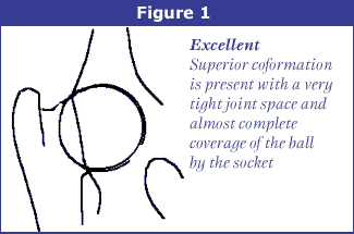
Excellent (Figure 1): this classification is assigned for
superior conformation in comparison to other animals of the same
age and breed. There is a deep seated ball (femoral head) which
fits tightly into a well-formed socket (acetabulum) with minimal
joint space. There is almost complete coverage of the socket over
the ball.
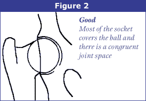
Good (Figure 2): slightly less than superior but a well-formed
congruent hip joint is visualized. The ball fits well into the
socket and good coverage is present.
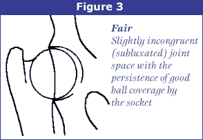
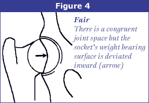
Fair (Figure 3): Assigned where minor irregularities in the hip
joint exist. The hip joint is wider than a good hip phenotype.
This is due to the ball slightly slipping out of the socket
causing a minor degree of joint incongruency. There may also be
slight inward deviation of the weight-bearing surface of the
socket (dorsal acetabular rim) causing the socket to appear
slightly shallow (Figure 4). This can be a normal finding in some
breeds however, such as the Chinese Shar Pei, Chow Chow, and
Poodle.
Borderline: there is no clear cut consensus between the
radiologists to place the hip into a given category of normal or
dysplastic. There is usually more incongruency present than what
occurs in the minor amount found in a fair but there are no
arthritic changes present that definitively diagnose the hip joint
being dysplastic. There also may be a bony projection present on
any of the areas of the hip anatomy illustrated above that can not
accurately be assessed as being an abnormal arthritic change or as
a normal anatomic variant for that individual dog. To increase the
accuracy of a correct diagnosis, it is recommended to repeat the
radiographs at a later date (usually 6 months). This allows the
radiologist to compare the initial film with the most recent film
over a given time period and assess for progressive arthritic
changes that would be expected if the dog was truly dysplastic.
Most dogs with this grade (over 50%) show no change in hip
conformation over time and receive a normal hip rating; usually a
fair hip phenotype.
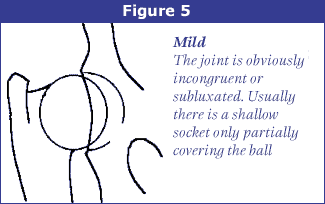
Mild Canine Hip Dysplasia (Figure 5): there is significant
subluxation present where the ball is partially out of the socket
causing an incongruent increased joint space. The socket is
usually shallow only partially covering the ball. There are
usually no arthritic changes present with this classification and
if the dog is young (24 to 30 months of age), there is an option
to resubmit an radiograph when the dog is older so it can be
reevaluated a second time. Most dogs will remain dysplastic
showing progression of the disease with early arthritic changes.
Since HD is a chronic, progressive disease, the older the dog, the
more accurate the diagnosis of HD (or lack of HD).
Moderate Canine Hip Dysplasia: there is significant subluxation
present where the ball is barely seated into a shallow socket
causing joint incongruency. There are secondary arthritic bone
changes usually along the femoral neck and head (termed remodeling),
acetabular rim changes (termed osteophytes or bone spurs) and
various degrees of trabecular bone pattern changes called
sclerosis. Once arthritis is reported, there is only continued
progression of arthritis over time.
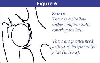
Severe HD (Figure 6): assigned where radiographic evidence of
marked dysplasia exists. There is significant subluxation present
where the ball is partly or completely out of a shallow socket.
Like moderate HD, there are also large amounts of secondary
arthritic bone changes along the femoral neck and head, acetabular
rim changes and large amounts of abnormal bone pattern changes.
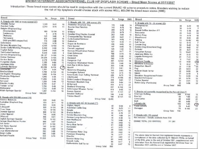
Other Hip Dysplasia Registries—An Approximation
The Lower the score the better the hips
|
|
FCI (European) |
BVA (UK/Australia) |
SV (Germany) |
|
E
|
A-1
|
0-4 (no > 3/hip)
|
Normal
|
|
G
|
A-2
|
5-10 (no > 6/hip)
|
Normal
|
|
F
|
B-1
|
11-18
|
Normal
|
|
B
|
B-2
|
19-25
|
Fast Normal
|
|
M
|
C
|
26-35
|
Noch Zugelassen
|
|
Mod
|
D
|
36-50
|
Mittlere
|
|
S
|
E
|
51-106
|
Schwere
|
Elbow
displasia |
 The
genetic disorder, prcd-PRA , causes cells in the retina
at the back of the eye to degenerate and die, even though the
cells seem to develop normally early in life. The “rod” cells
operate in low light levels and are the first to lose normal
function. Night blindness results. Then the “cone” cells
gradually lose their normal function in full light situations.
Most affected dogs will eventually be blind. Typically, the
clinical disease is recognized first in early adolescence or
early adulthood. Since age at onset of disease varies among
breeds, you should read specific information for your dog.
Diagnosis of retinal disease can be difficult. Conditions that
seem to be prcd-PRA might instead be another disease and
might not be inherited. OptiGen’s genetic test assists in making
the diagnosis. It’s important to remember that not all retinal
disease is PRA and not all PRA is the prcd form of PRA.
Annual eye exams by a veterinary ophthalmologist will build a
history of eye health that will help to diagnose disease.
The
genetic disorder, prcd-PRA , causes cells in the retina
at the back of the eye to degenerate and die, even though the
cells seem to develop normally early in life. The “rod” cells
operate in low light levels and are the first to lose normal
function. Night blindness results. Then the “cone” cells
gradually lose their normal function in full light situations.
Most affected dogs will eventually be blind. Typically, the
clinical disease is recognized first in early adolescence or
early adulthood. Since age at onset of disease varies among
breeds, you should read specific information for your dog.
Diagnosis of retinal disease can be difficult. Conditions that
seem to be prcd-PRA might instead be another disease and
might not be inherited. OptiGen’s genetic test assists in making
the diagnosis. It’s important to remember that not all retinal
disease is PRA and not all PRA is the prcd form of PRA.
Annual eye exams by a veterinary ophthalmologist will build a
history of eye health that will help to diagnose disease.
